Carl G. Lange: First proponent of the “low” vs.”high” road?

I finally got to reading Lange’s The Emotions: A Psychophysiological Study from 1885 (English 1922 translation of the German version) and found the most amazing thing: a dual-pathway, “low” and “high” road model of emotional processing:

From Lange (1885)

From Lange (1885). The “low road” goes from the eye to CO’ to CV — all subcortically. The “high road” goes up via cortex before coming down to CV.

The pathway from the eye to CO’ leads to CV rather directly. CV is a motor stage somewhere unspecified but in the brainstem. This applies to “simple” cases. But when a “mental process” is involved, cortical stages of vision and taste must be engaged, namely CO” and CG”. Now, the “impulses” get to CV from cortex!

I didn’t know about this passage, which I found rather stunning!

Here’s what Lange says himself: “… those emotions which are due to a simple sense impression, a loud noise, a beautiful color combination, etc., the path to the vasomotor center must be quite direct, and the cerebral mechanism but slightly complicated… The matter becomes somewhat more complicated when those affections are involved which are produced not by a simple impression upon some sense-organ, but by some ‘mental process,’ some memory or association of ideas, even if the latter be due to
sense-impression.”

Do you know of earlier formulations of this type of dual-route model? If so, let me know!

Understanding the function of brain regions and brain networks

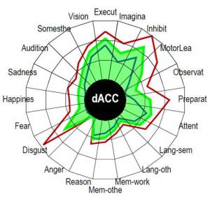
Michael Anderson, Josh Kinnison and I have just published a paper in Neuroimage describing a framework for capturing a brain region’s functional fingerprint, in addition to the fingerprint an entire network of brain regions. These are computed based on studies published in the BrainMap database and provide an estimate of the functional repertoire of a given brain region/network. For example, here’s the fingerprint of the dorsal ACC:

Functional fingerprint of the dorsal anterior cingulate cortex. The red and blue lines represent the uncertainty range of our estimate).

Functional fingerprint of the dorsal anterior cingulate cortex. The red and blue lines represent the uncertainty range of our estimate.

The framework allows the quantification of functional diversity (captured via Shannon’s entropy) in a voxelwise manner (by moving a 10-mm radius “spotlight” around). We found that most brain regions had diverse functional profiles, though diversity varied considerably across the brain (below, the zones in red are the most diverse):

Voxelwise functional diversity.

Voxelwise functional diversity.

The functional profiles of brain networks can also be characterized, such as the one of the “ventral attention network”:

Functional fingerprint of the ventral attention network.

Functional fingerprint of the ventral attention network.

Our study thus allowed us to characterize the contributions of individual brain regions and networks of brain regions without using singular task- or role-bound functional attributions.

Here’s the reference in case you have problems with the PDF link:

Anderson, M. L., Kinnison, J., & Pessoa, L. (2013). Describing functional diversity of brain regions and brain networks. NeuroImage, 73, 50–58.